How Cytology Centrifuge Smears are Prepared
Share

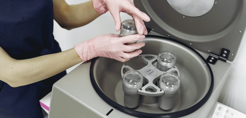
A cytology centrifuge is also known as cytocentrifuge. It is a specialized device that is used to concentrate the cells in fluid specimens onto a microscope slide. This is done so they can be stained and examined.
A cytology centrifuge is used in different areas of the clinical laboratory including microbiology, hematology, cytopathology, and biological research. The method can also be used in different types of specimens like urine, synovial fluid, and serous fluid.
Cytology Centrifuge Applications
Some of the common applications of cytocentrifuge include:
- Gram staining of fluid specimens (to identify microorganisms)
- Differential cell counts on body fluids (this includes cerebrospinal, synovial, and serous fluids)
- Cytopathology examination of liquid specimens (this includes body fluids and fine needle aspirates)

How Cytocentrifuge Smears are Prepared
When preparing a cytocentrifuge smear, a funnel assembly will be attached to the front of the microscope slide. The surface of the funnel assembly that is in contact with the slide will be lined with filter paper. This is done to absorb any excess fluid.
Few drops of sample will be placed in the funnel. The assembly will be placed in the cytocentrifuge (which operates at low force to preserve cellular structure). Centrifugal force will push the fluid through the opening of the funnel.
It also concentrates the cells in a small area. Centrifugation will concentrate cells by about twenty-fold. It also allows the creation of a one-cell-thick monolayer that can be used to assess cellular morphology.


Social Media