

The purpose of a cytology centrifuge is to concentrate cells in fluid specimens onto the microscope slide. The slides would be stained and analyzed for diagnostic or research purposes.
Preparation Procedure
A certain procedure is used when using a cytology centrifuge. The first thing you should do is attach a funnel assembly to the front of the microscope slide. The surface assembly of the funnel that is attached to the slide is also lined with filter paper. This is done so any excess liquid of the smear is easily absorbed.
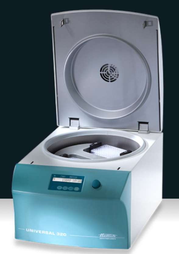
After samples have been placed in the funnel, the assembly is placed into the centrifuge before it is run at 600-800 x g. Using low force is also essential so the structure of the specimen is preserved once the centrifuge has been operated.
How Centrifugal Force is Used
Centrifugal force will force the fluid into the assembly so it goes toward the opening of the funnel and concentrates the cells in one area of the slide. The cells concentrated onto the slides. From there, it creates a one-cell-thick monolayer that makes cellular morphology assessment.
Where Cytology Centrifuge are Used
A cytology centrifuge is usually used in different diagnostic procedures, including:
- Gram staining of fluid specimens to check the presence of microorganisms
- Assessing of differential cell counts on body fluids, including serous, cerebrospinal, and synovial fluids
- Examination of liquid specimens such as fine needle aspirates and body fluids in cytopathology
Since cells are placed in the center of the fluid smear on the slide, they can appear compressed compared to those at the periphery that results from the centrifugation process.
To minimize overcrowding effects on the slide, specimens with a higher cell count need to be diluted before the sample is prepared.


